Home

Cercle de roulement audition Amplifier ecodoppler inguinale indice Tofu Labe

usg-16054-f3.gif
usg-16054-f3.gif

Radiologia Brasileira - Avaliação ultrassonográfica da dor inguinoescrotal:  uma revisão baseada em imagens para o ultrassonografista
Radiologia Brasileira - Avaliação ultrassonográfica da dor inguinoescrotal: uma revisão baseada em imagens para o ultrassonografista

Inguinal Hernia during Pregnancy- What you should know!

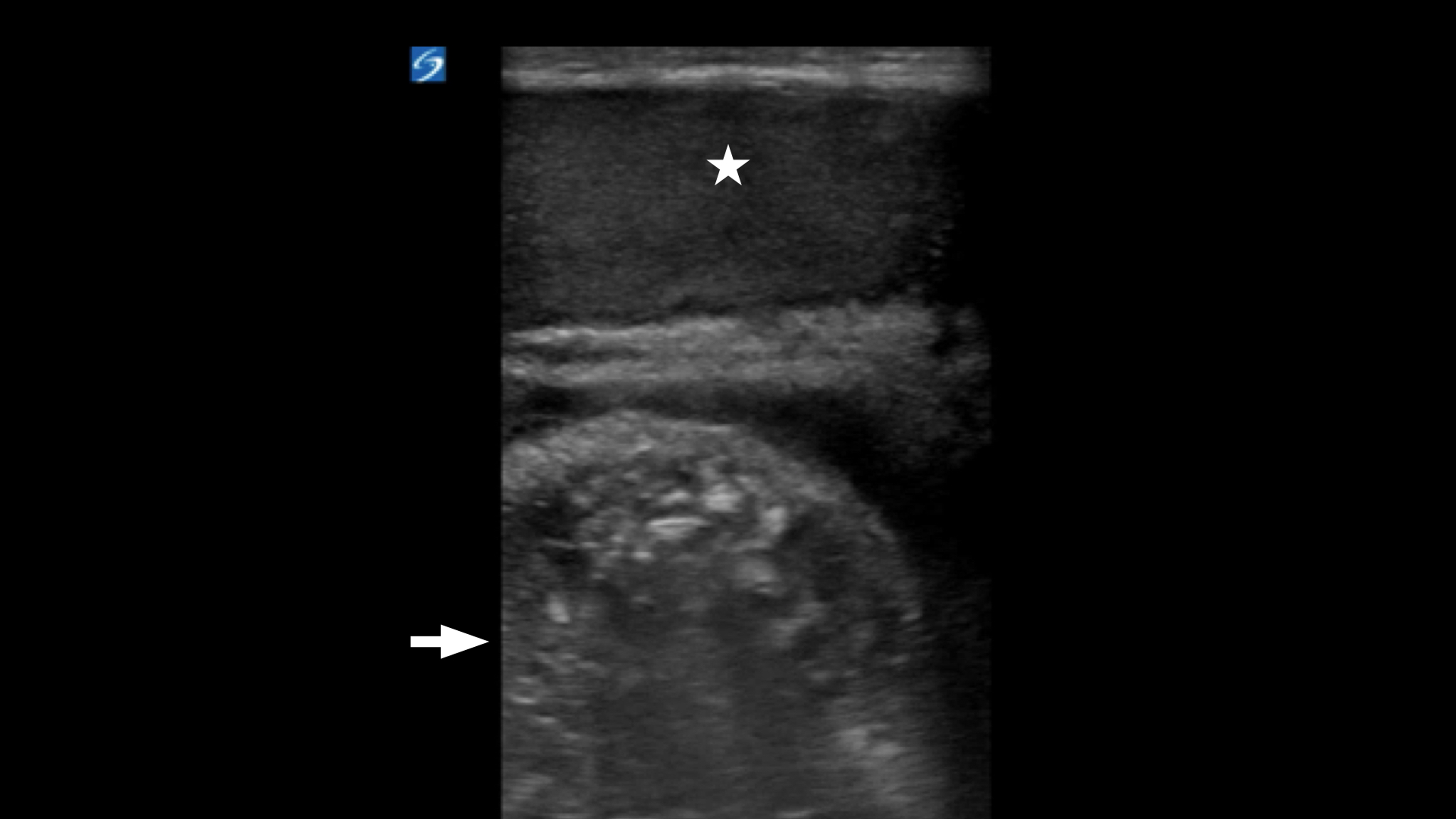
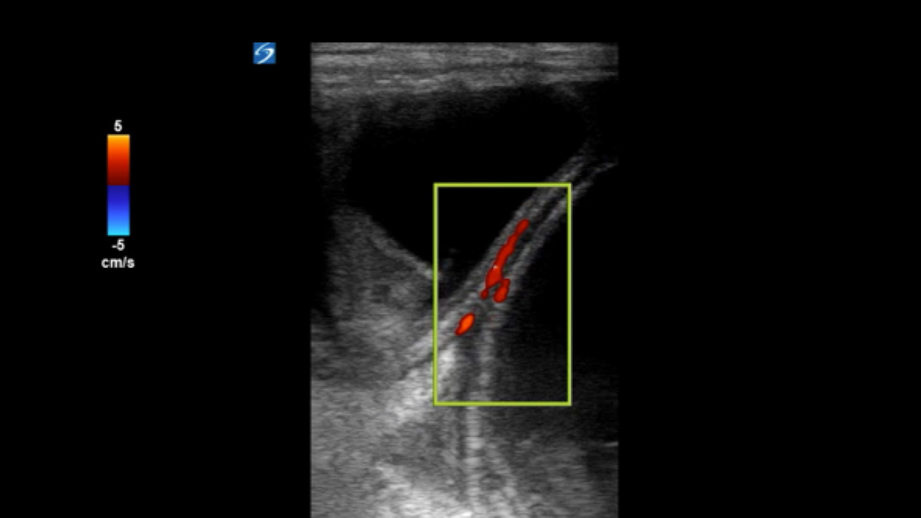
Cureus | Point-of-Care Ultrasound in the Diagnosis of an Incarcerated  Inguinal Hernia | Article
Cureus | Point-of-Care Ultrasound in the Diagnosis of an Incarcerated Inguinal Hernia | Article

Cureus | Point-of-Care Ultrasound in the Diagnosis of an Incarcerated  Inguinal Hernia | Article
Cureus | Point-of-Care Ultrasound in the Diagnosis of an Incarcerated Inguinal Hernia | Article

Figure 1 from Round ligament varicosities mimicking inguinal hernias in  pregnancy: importance of color Doppler sonography. | Semantic Scholar
Figure 1 from Round ligament varicosities mimicking inguinal hernias in pregnancy: importance of color Doppler sonography. | Semantic Scholar

Enlarged inguinal lymph node (arrows) demonstrating flow in the hilar... |  Download Scientific Diagram
Enlarged inguinal lymph node (arrows) demonstrating flow in the hilar... | Download Scientific Diagram
Cryptorchidism - Wikipedia

Ultrasonography for lymph nodes metastasis identification in bitches with  mammary neoplasms | Scientific Reports
Ultrasonography for lymph nodes metastasis identification in bitches with mammary neoplasms | Scientific Reports

PDF] Incarcerated Inguinal Hernia: A Cause of Testicular Ischemia Without  the 'Twist' | Semantic Scholar
PDF] Incarcerated Inguinal Hernia: A Cause of Testicular Ischemia Without the 'Twist' | Semantic Scholar

Figure 1. (a) Doppler ultrasonography (DU) shows a well-defined, echo-free,  cystic mass in the right inguinal region. (b) Color doppler flow image  shows that the cystic mass obviously compresses the common femoral
Figure 1. (a) Doppler ultrasonography (DU) shows a well-defined, echo-free, cystic mass in the right inguinal region. (b) Color doppler flow image shows that the cystic mass obviously compresses the common femoral

EPOS™
EPOS™

a-e trans-perineal and inguinal ultrasound and color Doppler images... |  Download Scientific Diagram
a-e trans-perineal and inguinal ultrasound and color Doppler images... | Download Scientific Diagram

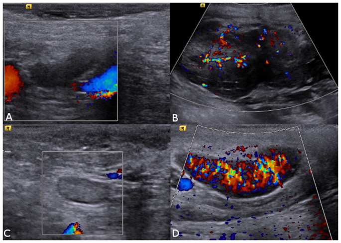
Ultrasound Doppler of right inguinal mass showing Multiple nodular... |  Download Scientific Diagram
Ultrasound Doppler of right inguinal mass showing Multiple nodular... | Download Scientific Diagram

Testicular Compromise due to Inguinal Hernia - The Western Journal of  Emergency Medicine
Testicular Compromise due to Inguinal Hernia - The Western Journal of Emergency Medicine

Testicular Torsion | UAMS Department of Radiology

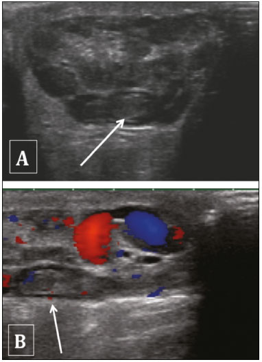
Fat-containing inguinal hernia. Gray-scale (A) and color Doppler (B) US...  | Download Scientific Diagram
Fat-containing inguinal hernia. Gray-scale (A) and color Doppler (B) US... | Download Scientific Diagram

Grayscale ultrasound and color Doppler ultrasound imaging of the right... |  Download Scientific Diagram
Grayscale ultrasound and color Doppler ultrasound imaging of the right... | Download Scientific Diagram